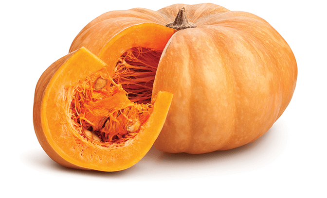
 We’re often most frightened of those things we can’t see or understand. Fortunately optical coherence tomography gives us ‘eyes’ to see below the surface, revealing detailed structure at the micron level through non-contact, nondestructive imaging. This technology can be applied to tissue in the body as easily as plant tissue (like pumpkin), and can be used for research, diagnostics, or even quality control.
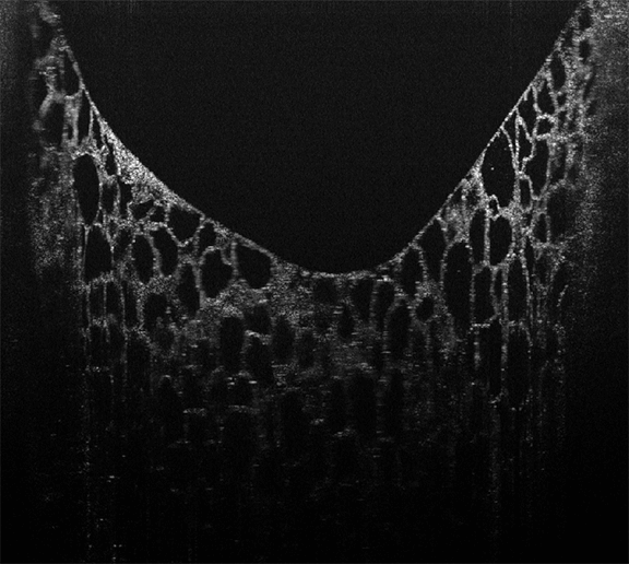
OCT provides cross-sectional images 1-10 mm deep into materials, which can be combined to create 3D volume views, or to see an x-y plane at a specified depth in a sample. This OCT image taken of pumpkin rind from the inside in cross-section shows very clearly the large cells that make up its tissue. It was taken with an OCT system designed using one of our high-resolution, high-speed Cobra-S 800 nm OCT spectrometers.
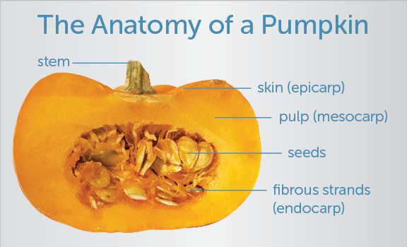
See how the fibers mysteriously appear and disappear in the video below? We’ve re-analyzed a 3D section of pumpkin tissue to give a top-down (eg., en face) view, zooming from the exposed surface into its depths to show the fibers of vascular tissue that pervade the mesocarp, the fleshy tissue that makes up a pumpkin’s shell (explore the pumpkin’s fascinating anatomy from a practical viewpoint here).
To see more of the amazing images that can be generated using OCT, explore our image gallery. Or read our OCT tutorial to demystify the science behind this imaging technology!
