 Ophthalmic imaging is an integral part of diagnostic procedures evaluating eye conditions. It enables ophthalmologists to image actual living tissue deep in the human eye at microscopic resolution and thus provides the basis for treatments of eye diseases.
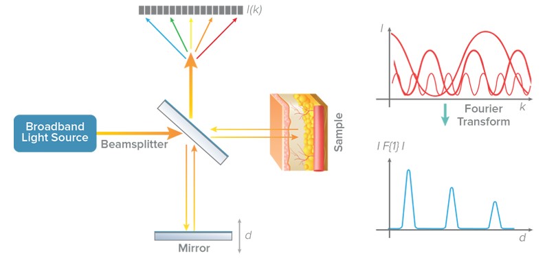
Optical coherence tomography, or OCT, is the method of choice for ophthalmologists looking to examine the back of the eye, known as the retina, where standard techniques reach their limits. This imaging technique uses infrared light to probe the eye and creates a 3D image from a series of cross sections on a very short time scale.
Spectral-domain optical coherence tomography is a subset of OCT technology that combines a light source with broad optical bandwidth and a high sensitivity spectrometer with high signal-to-noise-ratio for detection. It gains its name from the spectra of back-scattered light that are collected from different depths within a sample to generate the OCT image.
A High-Resolution Look into the Eye
The eye is a very sensitive and complex organ that consists of several different intricate structures. Spectral-domain optical coherence tomography, often abbreviated to SD-OCT, therefore provides a unique method for ophthalmologists to observe retinal structure that is key to the diagnosis of injury and disease during eye examinations.
Key advantages of SD-OCT include the non-contact and non-destructive approach to observing the eye, a very high imaging resolution, and its ability to be performed quickly, without any special patient prep. It is perhaps the easiest medical imaging test that a patient can take and it only lasts about 5 minutes.
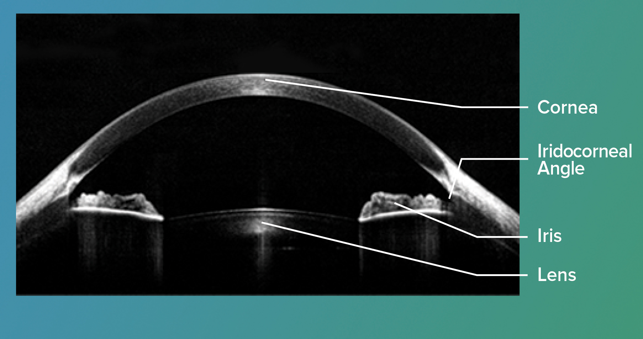
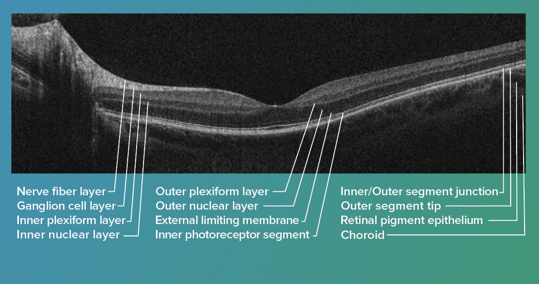
Ophthalmologists commonly divide the eye into two parts: the front of the eye, known as the anterior segment, and the area behind the lens, the posterior segment. Imaging these two segments with spectral-domain optical coherence tomography requires different wavelengths in the infrared region to probe the eye perfectly. For example, the cornea, part of the anterior segment, is examined with 800 nm wavelength in proximity to the surface while 1300 nm wavelength is suitable for deeper anterior structures such as the lens. Examination of the anterior segment is crucial for treatments of diseases such as glaucoma and cataracts. The main parts of the posterior segment are the retina, macula, and optic nerve. By means of spectral-domain optical coherence tomography, high resolution details of the retina may also be obtained with 800 nm wavelength, while a wavelength of 1050 nm is suitable for examinations of the optic nerve. Examining the posterior region is relevant in the diagnostic of age-related macular degeneration (AMD) which is a leading cause of vision loss in elderly people.
 Ultra hi-resolution SD-OCT imaging of the cornea is being explored by Dr. Kostadinka Bizheva at the University of Waterloo as a future method to advance disease diagnosis & treatment.
See the detailed images they were able to capture in just 2.8 seconds.
SD-OCT Gratings, Spectrometers and SDKs
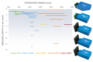 Wasatch Photonics offer high performance gratings, spectrometers and OEM modules for spectral-domain optical coherence tomography. In fact, our VPH gratings are the industry-leading grating for SD-OCT instruments, and are deployed around the world. We also offer the Cobra OCT spectrometer series to speed OCT system development, at a variety of bandwidths and scan rates centered on the key OCT wavelengths of 550, 800, 1050, 1300, and 1600 nm. Our spectrometer design is based on our own in-house VPH transmission gratings and optimized optics to deliver high efficiency and low-roll off for the clearest, deepest images.
If you are looking to speed development of your own system, we can provide drop-in OEM solutions at the grating, spectrometer, and module levels customized to your individual needs for spectral-domain optical coherence tomography. We work with the best cameras on the market and support you with our OCT Software Development Kits for simplified and easy control of our standard and custom OEM OCT spectrometers.
Contact us directly to start a discussion today.